Leading Technology
All our Clinics are fully equipped with the most up to date technology including ophthalmic diagnostic equipment, assisting our doctors to accurately diagnose and deliver the appropriate patient treatment.
Optical Coherence Tomography (OCT)
An OCT image is a vital part of the assessment of your macula and retina and especially useful in the diagnosis and management of conditions such as age-related macular degeneration (AMD), epiretinal membranes (ERM), macular hole, central serous chorioretinopathy (CSCR), diabetic eye disease, vitreomacular traction (VMT) and vascular problems of the eye.
OCT imaging is non invasive and painless and results are available immediately. It provides a detailed cross-sectional picture of the retinas structure at a microscopic level. The routine use of OCT imaging has meant that more invasive and expensive procedures are sometimes not needed.
The state of the art Heidlberg Spectralis used in our practices is able to scan the retina in intricate detail. The OCT camera uses a very accurate low power laser that scans rapidly across the macula to give an image much like an ultrasound. As the wavelength of light is so much smaller than that of sound waves, the OCT image of the retina is exceptionally detailed and allows accurate measurements of retinal thickness.
An OCT will be performed on most visits to the clinic. It allows the doctor to compare the appearance and measure changes at the macula, this is particularly important for patients receiving treatments, such as intravitreal injections.
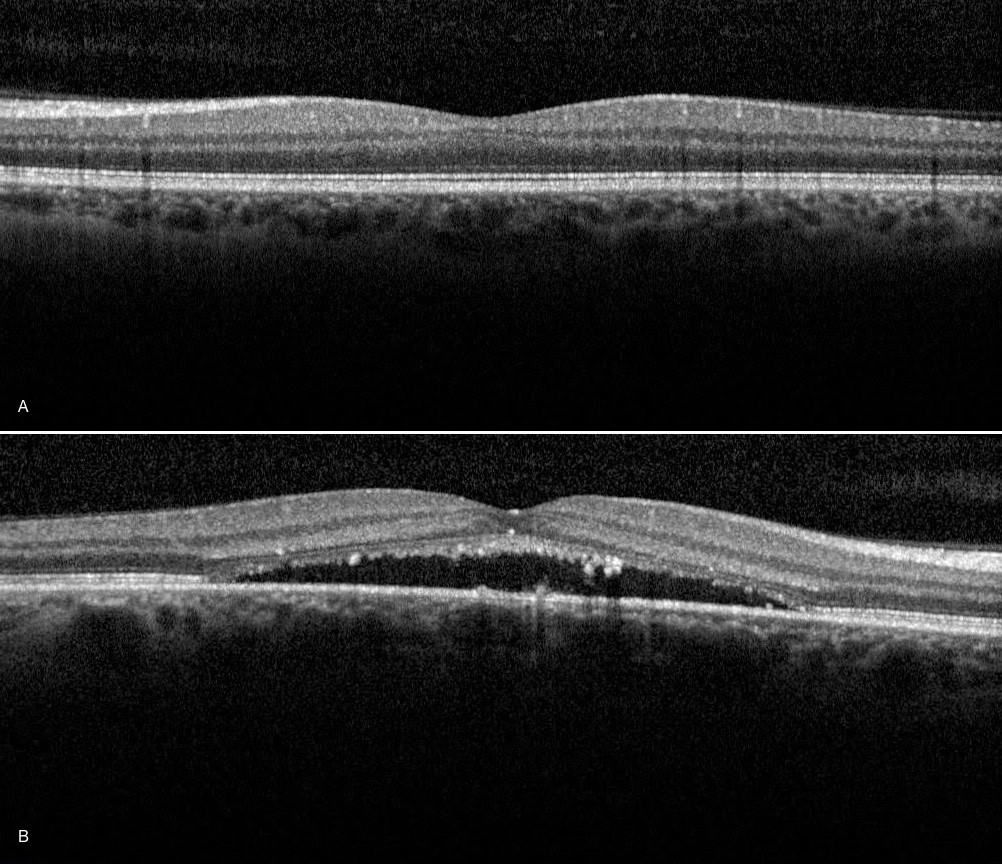
Image A: OCT of normal macula. Image B: OCT of abnormal macula
Fluorescein Angiography (FA)
A fluorescein angiogram (FA) is a special photographic test that is used to evaluate the retinal circulation and other retinal structures in serious retinal and macular conditions.
It is not an X RAY. This test is most often used in the management of age related macular degeneration, diabetic eye disease and retinal vascular occlusions.
At Retina Consultants we use the Heidlberg Spectralis machine (which also collects OCT images) for FA. This allows extremely accurate images to be collected using a minimum of light; which makes the test more comfortable and a minimal amount of fluorescein dye (which makes the test safer).
During a FA a yellow vegetable dye called fluorescein is injected into a vein in the back of the hand or arm. The dye travels quickly via the blood stream to the eye. Multiple photographs are taken using a special camera. The test demonstrates in minute detail abnormalities of the retinal blood structures and vessels and helps in the diagnosis of many retinal conditions.
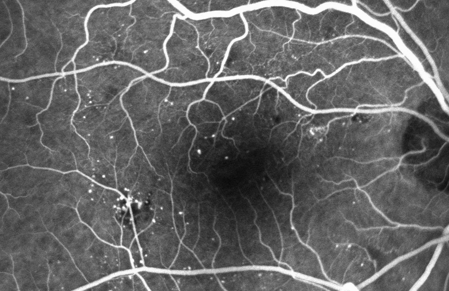
Fluorescein Angiogram of a diabetic retina
Indocyanine Green Angiography (ICG)
This is a photographic test similar to a Fluorescein Angiography (FA) except that a special dye called Indocyanine Green (ICG) is used. Often an ICG is done in tandem with a fluorescein angiogram. The ICG dye allows better visualisation of the choroidal circulation, which is the vascular layer located under the retina, which is otherwise difficult to see. This gives helpful information in unusual cases of macular degeneration, particularly when associated with haemorrhage
ICG is particularly useful in studying the choroidal vasculature and circulation in the eyes of patients with Chronic Serous Chorioretinopathy (CSCR) and in some cases of wet age related macular degeneration.
ICG is an Iodine based dye, which is injected into a vein in the arm or back of hand, where it travels via the blood stream to the eye. Photos of the blood vessels of the eye are taken by the Spectralis machine. The dye becomes visible when excited by infrared light.
The test takes 20 minutes to perform. As ICG is Iodine based, it is not used if you are allergic to Iodine or seafood.
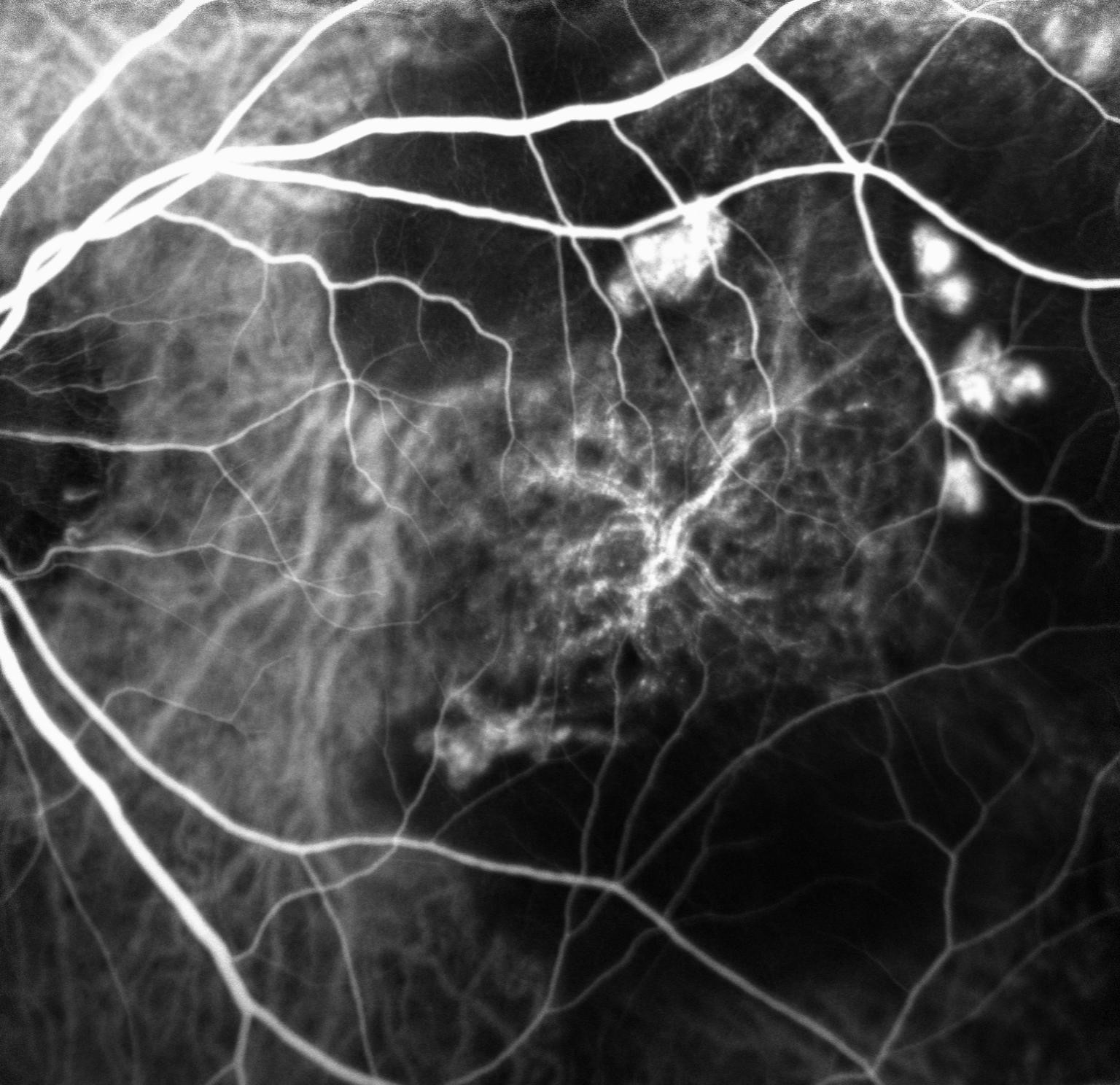
ICG angiogram showing Retinal Polyps
Digital Retinal Photography
Digital photography is used to take high resolution photos of the retina and macula.
These high definition photos show fine details of the retina and retinal pathology allowing the Ophthalmologists to demonstrate to the patient their retinal condition.
These images are often captured on an initial consultation and used as a baseline to compare and monitor a person’s condition over time.
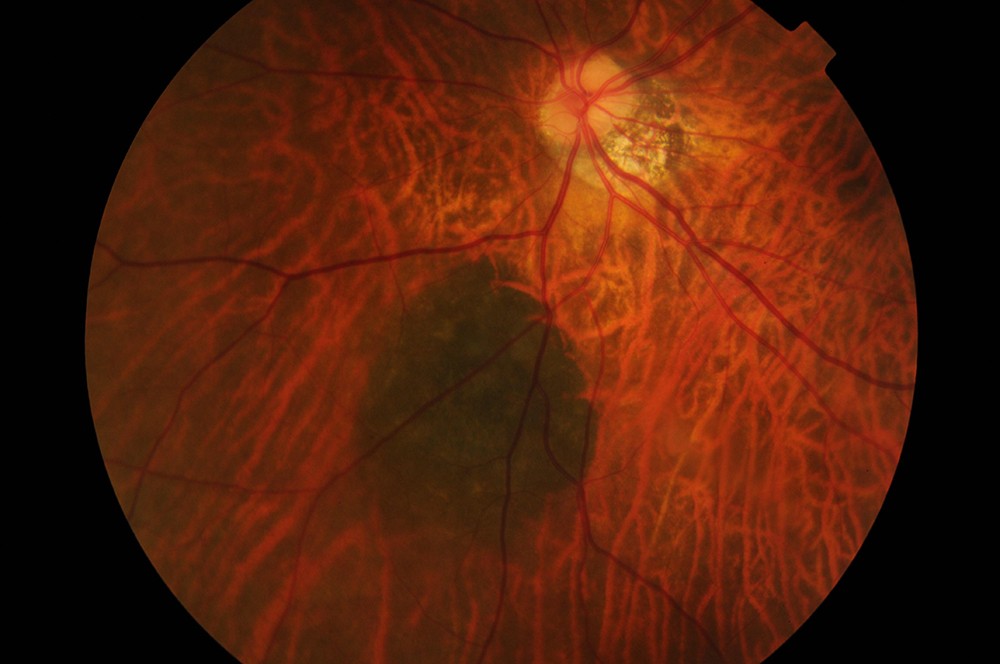
Digital retinal photograh of a retinal naevus
Ocular Ultrasound (B SCAN)
A B scan is an ultrasound of the internal structures of the eye. It is performed when the retina is unable to be visualised, due to dense cataract (opacity of the lens of the eye) or opacity of the vitreous jelly for example in the case of vitreous haemorrhage. It is most frequently used in the case of retinal detachment or used to characterise and measure the size of a mass in the eye, such as a choroidal naevus or melanoma.
B scans are painless and non invasive. A gel is applied over the closed eyelid and a special probe is rolled over the eyelid. The ultrasound uses sound waves to produce an image, just like an ultrasound of an unborn baby. It takes five minutes and results are available immediately
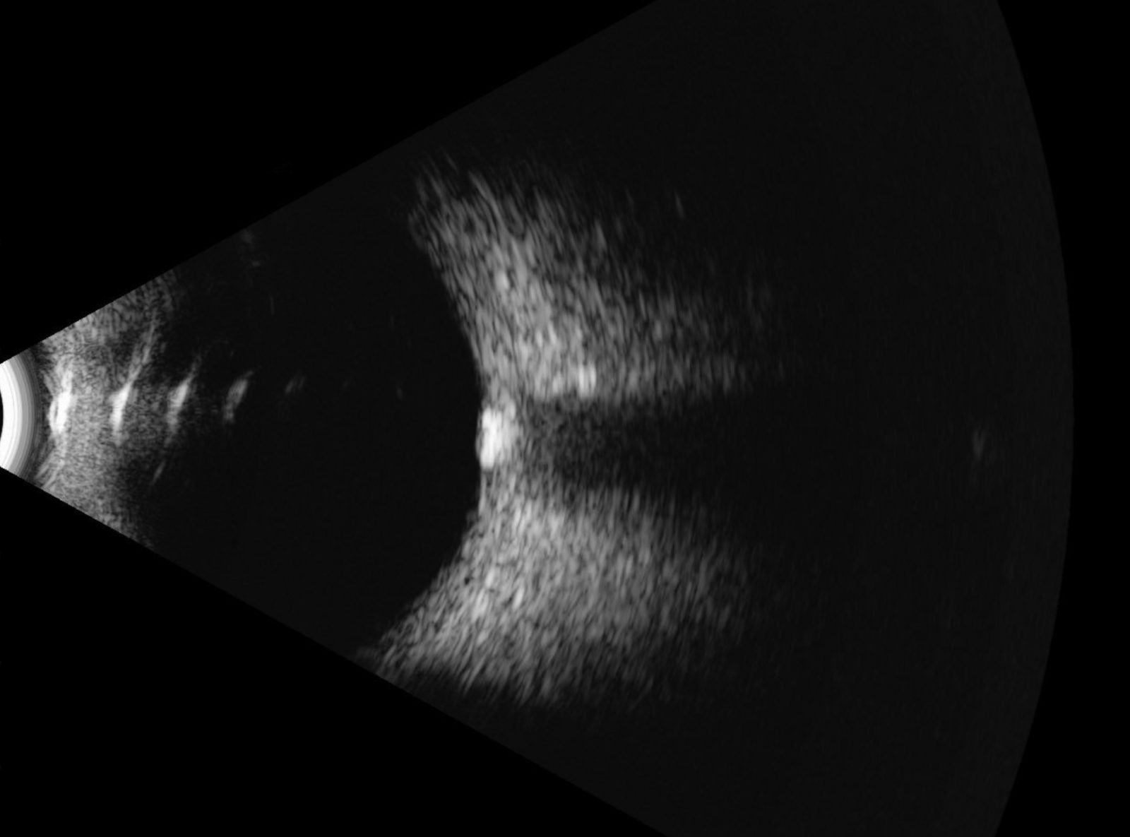
B-scan of the eye